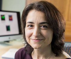
Mapping glioblastoma cells
Fast facts
- Official title: Mapping the spatio-temporal heterogeneity of glioblastoma invasion
- Lead researcher: Professor Simona Parrinello
- Where: University College London
- When: September 2019 – February 2025
- Cost: £1,496,690 (supported by The Oli Hilsdon Foundation)
- Research type: Academic, Lab-based, Glioblastoma, Adult
- Grant round: Quest for Cures
What is it?
Glioblastomas are diffuse in nature, meaning the tumour cells can spread through healthy parts of the brain. This is through a process called invasion, and makes it difficult to completely remove the tumour during surgery. Due to this, recurrence is a risk with glioblastomas as the tumour cells left behind can grow into a new tumour.
Professor Parrinello, and her colleagues at Imperial College London, aim to understand how glioblastomas spread into the brain and how they use small molecules as messengers to communicate with surrounding cells. It is likely that glioblastoma use different molecules to communicate with different types of cells, which makes identifying the various molecules difficult.
The multi-disciplinary research team will investigate this communication by combining both biological and mathematical principles and using two new techniques called spatial transcriptomics and intravital photoacoustic imaging (PAI).
Spatial transcriptomics is a technique that allows researchers to characterise different cells while preserving information about the cells’ original location.
Characterisation of the cells means that we will know what changes are present in cells at each location. If the changes vary in different areas of the tumour it gives us insight into how to develop more specific treatments.
PAI is an advanced imaging technique that uses light and sound to create high resolution images. The research team will use PAI to create images of the brain that will help them visualise the invasion of glioblastoma cells deep inside the brain.
These two techniques will allow the researchers to map the invasion process and identify key molecules that help the tumour cells spread. This understanding will help researchers develop more effective therapies that will help block tumour cells from spreading and prevent recurrence.
Why it is important?
Currently, not much is known about how a glioblastoma invades the surrounding brain because:
- tumour cells that spread deep into the brain cannot be studied easily
- there are considerable differences within a single tumour and between each person
- the brain is complex and contains many different types of cells in different regions of the brain.
These obstacles are some of the reasons that, even with the current gold standard of care, a glioblastoma diagnosis comes with a poor prognosis and, even after treatment, tumour recurrence is very likely.
This research project aims to address all these obstacles and help us find drug targets to prevent tumour recurrence. Giving people with a glioblastoma much-needed hope for a future where brain tumours are defeated.
Who it will help?
The researchers on this project have a broad range of interests and are coming together to fill a significant gap in our knowledge, namely ‘why do glioblastomas keep coming back?’
This work has the potential to benefit everyone who has had treatment for a glioblastoma and is living with the worry that it will return.
Milestones
-
- Generated 20 tumours derived from patient cells. 15 were used to produce tumours in preclinical models which can now be analysed with spatial transcriptomics, imaging and computer analysis.
- Set up the experiments to start carrying out spatial transcriptomics on different tumour cells and have generated their first data set.
- Developed methods for analysing the data from spatial transcriptomics with computer analysis.
- Set up imaging methods for determining tumour cell location and brain anatomy in the samples. This includes MRI, to track tumour growth in real time.
-
- Are now able to distinguish between normal cells and tumour cells in spatial transcriptomic data using computer methods.
- Found genes that can be used to identify changes in GBM cell behaviour during invasion.
- Worked on imaging methods for determining tumour cell location and brain anatomy including one that could be readily translated to a clinical setting.
- Developed mathematical methods for simulating basic aspects of GBM as well as statistical methods to compare these virtual models with real-life data.
-
- Identified one of the key molecules responsible for the invasive nature of GBM, SOX9.
- Found two patterns that are used by GBM to infiltrate the brain, each method depends on the tumours genetic makeup.
- Currently they are setting up experiments to study how invasion and SOX9 works.
-
- Found that GBMs grow due to the damaged, surrounding normal brain cells perpetuating it. This occurs specifically in the white matter of the brain, where GBMs tend to develop.
- This response is caused by the injured neurons triggering Wallerian degeneration which accelerates tumour growth. Blocking it causes tumour growth to slow down.
- Identified another molecule (Pdgfa) that is activated by dying neurons and fuels tumour growth.
If you have any questions about this, or any of our other research projects, please contact us on research@thebraintumourcharity.org
Research is just one other way your regular gift can make a difference
Research is the only way we will discover kinder, more effective treatments and, ultimately, stamp out brain tumours – for good! However, brain tumours are complex and research in to them takes a great deal of time and money.
Across the UK, over 100,000 families are facing the overwhelming diagnosis of a brain tumour and it is only through the generosity of people like you can we continue to help them.
But, by setting up a regular gift – as little as £2 per month – you can ensure that families no longer face this destructive disease.
In this section

Professor Simona Parrinello
Prof Parrinello is a professor of neuro-oncology at University College London in the department of Cancer Biology.
